
Print
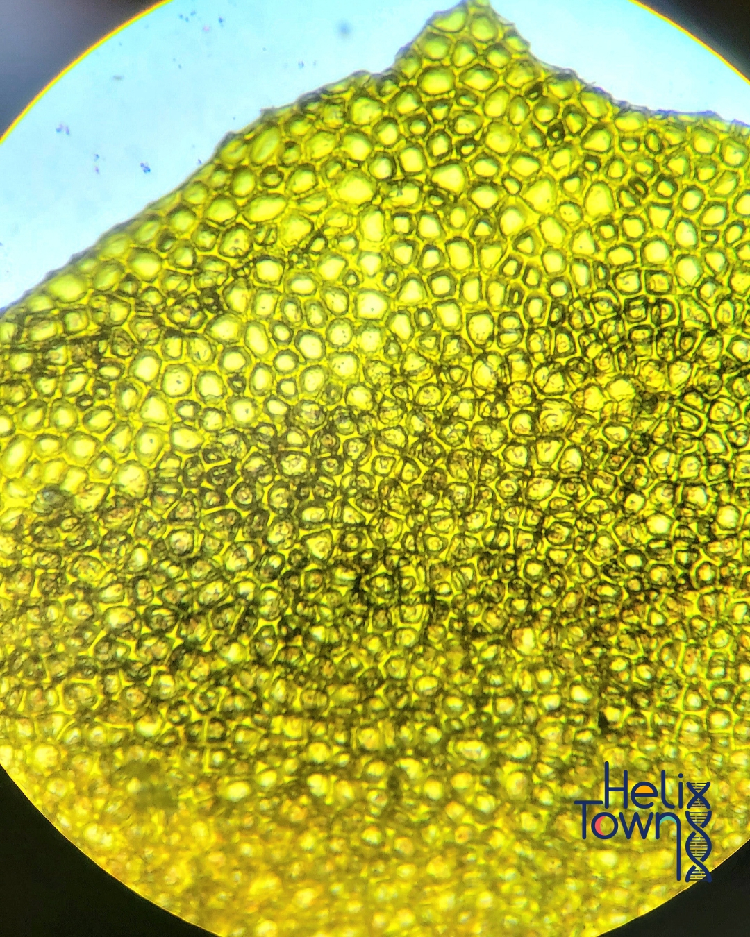
Explore the microscopic world inside a tomato by observing its cells under a microscope. Learn how plant cells are organized and identify basic structures such as the cell wall and cytoplasm.
Materials
- 1 small ripe tomato
- Tweezers
- Microscope
- Glass slide and coverslip
- Dropper or pipette
- Paper towel
Instructions
- Place one small drop of distilled water (dH₂O) in the center of a clean glass slide. This keeps the peel hydrated and easy to flatten.
- Use tweezers to gently lift a tiny, transparent piece of tomato skin from the outer surface of the tomato.Choose a piece that is as thin as possible (the inner film of the peel works best).Avoid thick or juicy parts of the flesh.
- Lay the peel flat on the drop of water. Smooth gently with tweezers to remove folds or bubbles.
- Hold the coverslip at a 45° angle and lower it slowly over the sample to avoid trapping air bubbles.
- Start with low power to find the cells. Switch to high power for details.
- Draw or photograph your view. Label cell wall and cells.Cell wall: clear or slightly outlined rectangular or irregular borders.
What's going on?
- The tomato peel is made of epidermal plant cells that protect the fruit.
- The cell wall is a defining feature of all plant cells and gives each cell its distinct shape.
Level
- Intermediate
Tried this experiment?Please consider Leaving a Review!