
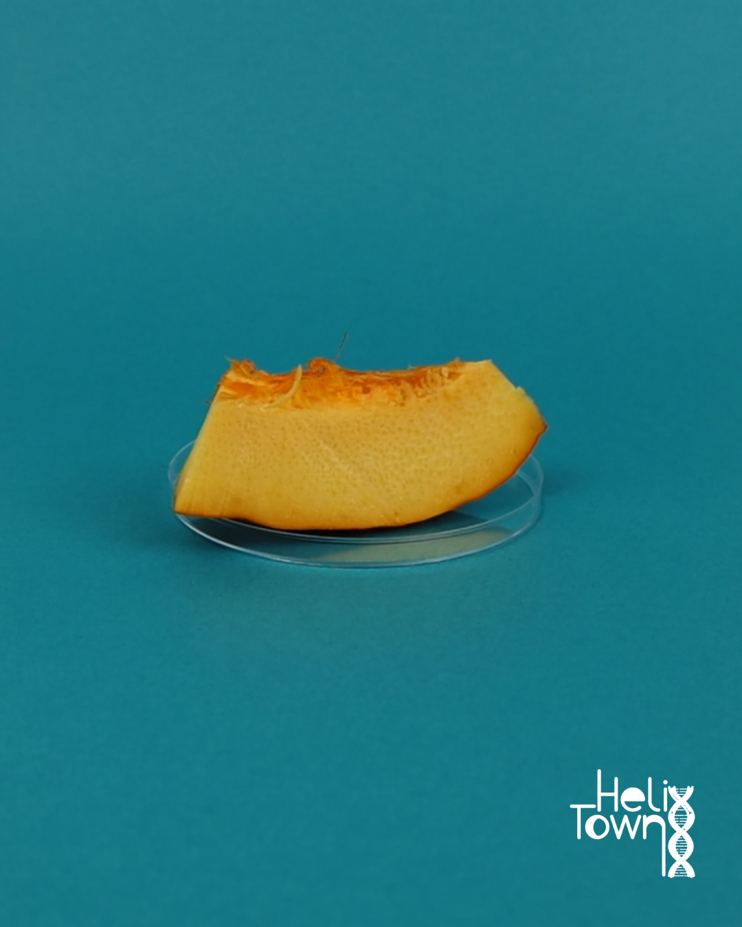
Materials
- Fresh pumpkin piece (orange outer skin)
- Sharp blade or scalpel (adult supervision required)
- Tweezers
- Microscope
- Glass slide and coverslip
- Dropper or pipette
- Distilled water
Instructions
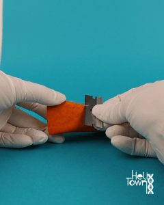
- With adult supervision, cut a very thin layer from the orange surface of the pumpkin skin (the outermost layer).
- Place one small drop of water in the center of a clean glass slide.
- Lay the thin slice of pumpkin skin flat on the water drop.Use tweezers to smooth it out so it lies flat without folds.

- Lower a coverslip gently at a 45° angle to avoid trapping air bubbles.

- Start at low power to locate the epidermal cells.
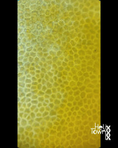
- Switch to high power to find stomata (tiny openings) surrounded by guard cells (two curved, bean-shaped cells).

- Draw or photograph your field of view. Label stoma, guard cells and epidermal cells.
What’s going on?
- The epidermis of the pumpkin skin forms a protective outer layer that prevents water loss.
- The stomata are small openings that allow gases to move in and out.
- Each stoma is surrounded by two guard cells that open and close the pore to regulate airflow.
- These features help the pumpkin exchange gases and control water balance, just like leaves do!
- Fun Fact: The word "stoma" originates from the Greek word for "mouth." The plural of "stoma" is "stomata.
Tried this experiment?Please consider Leaving a Review!